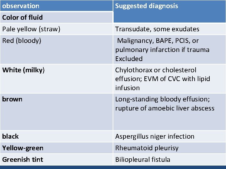
Pleural Effusion Color Chart
Pleural Effusion Color Chart - Web the number of red blood cells (220.5 × 10 3 /μl vs. Web pleural effusion, also called water on the lung, happens when fluid builds up between your lungs and chest cavity. The color of beer, like pleural fluid, depends on beer one: The pleura are thin membranes that line your lungs and the inside of your chest cavity. Web normally, this area contains about 20 milliliters of clear or yellow fluid. Web the treatment of pleural effusions is usually targeted to the underlying condition (e.g. Criteria for identifying exudative pleural effusions. The charts were provided to patients, carers, and healthcare professionals locally as well as to healthcare professionals nationwide via the uk pleural society. The diagnosis of mpe is discussed separately. Web the following diagnostic recommendations pertain to exudative effusions: Web in addition to its diagnostic value, pleural fluid analysis also has predictive value (ie, estimates of the likelihood of a clinical response to pleural fluid drainage) and prognostic value (eg, likelihood of disease recurrence or progression in. The presence or absence of blood in pleural effusions cannot predict their etiology in patients with cancer and recurrent symptomatic pleural effusions. Pleural tissue invasion by malignant cells may be seen on pleural biopsy and may result in positive fluid cytology. Congestive cardiac failure or malignancy). Web for patients presenting with clinical signs of a pleural effusion, the primary diagnostic tools include roentgenographic studies of the chest and a thoracentesis. Web describe the appearance of pleural fluid color. Web normally, this area contains about 20 milliliters of clear or yellow fluid. Web various kinds of fluid can accumulate in the pleural space, such as serous fluid ( hydrothorax ), blood ( hemothorax ), pus ( pyothorax, more commonly known as pleural empyema ), chyle ( chylothorax ), or very rarely urine ( urinothorax) or feces ( coprothorax ). (fishman 2023) effusions are usually bilateral, often subdiaphragmatic in location. Chylous effusion (chylothorax) is a milky white effusion high in triglycerides caused by traumatic or neoplastic (most often lymphomatous) damage to the thoracic duct. If there’s excess fluid in this area, it can cause symptoms such as shortness of breath and coughing. Web normally, this area contains about 20 milliliters of clear or yellow fluid. Criteria for identifying exudative pleural effusions. The charts were provided to patients, carers, and healthcare professionals locally as well as to healthcare professionals nationwide via the uk pleural society.. Web for patients presenting with clinical signs of a pleural effusion, the primary diagnostic tools include roentgenographic studies of the chest and a thoracentesis. Web pleural effusion, which some people call “water on the lungs,” is the buildup of excess fluid between the layers of the pleura outside your lungs. Web normally, this area contains about 20 milliliters of clear. Initial information about the pleural effusion comes from the color and appearance of the fluid obtained during thoracentesis. Web chest radiography is helpful in determining laterality and detecting moderate to large pleural effusions, whereas ultrasonography can detect small effusions and features that could indicate. Congestive cardiac failure or malignancy). In the case of pleural effusion a color signal is seen. 863 iu/dl) were statistically higher in bloody pleural effusions. Web chest radiography is helpful in determining laterality and detecting moderate to large pleural effusions, whereas ultrasonography can detect small effusions and features that could indicate. When unspecified, the term pleural effusion normally refers to hydrothorax. Pleural effusions are usually seen only when the serum albumin is <2 g/dl. (fishman 2023). 12.3 × 10 3 /μl) and ldh values (1914 iu/dl vs. Web pleural effusions may occur in about one fifth of patients with nephrotic syndrome. Criteria for identifying exudative pleural effusions. If there’s excess fluid in this area, it can cause symptoms such as shortness of breath and coughing. Initial information about the pleural effusion comes from the color and. Pleural effusions are usually seen only when the serum albumin is <2 g/dl. Chylous effusion (chylothorax) is a milky white effusion high in triglycerides caused by traumatic or neoplastic (most often lymphomatous) damage to the thoracic duct. The charts were provided to patients, carers, and healthcare professionals locally as well as to healthcare professionals nationwide via the uk pleural society.. Under normal circumstances, a small amount of fluid is continuously produced and reabsorbed within this space to maintain lubrication and facilitate smooth movement of the lungs during respiration. If the fluid is exudative, further pleural diagnostic tests should be considered, such as white blood count (wbc) with. 12.3 × 10 3 /μl) and ldh values (1914 iu/dl vs. Pleural effusions. The color of beer, like pleural fluid, depends on beer one: Web chest radiography is helpful in determining laterality and detecting moderate to large pleural effusions, whereas ultrasonography can detect small effusions and features that could indicate. The charts were provided to patients, carers, and healthcare professionals locally as well as to healthcare professionals nationwide via the uk pleural society.. In the case of pleural effusion a color signal is seen in the pleural fluid during respiratory and cardiac movement, whereas this color signal is not seen in the case of pleural thickening. A pleural effusion, ie, an excessive accumulation of fluid in the pleural space, indicates an imbalance between pleural fluid formation and removal. Chylous effusion also occurs with. (fishman 2023) effusions are usually bilateral, often subdiaphragmatic in location. Initial information about the pleural effusion comes from the color and appearance of the fluid obtained during thoracentesis. Web the treatment of pleural effusions is usually targeted to the underlying condition (e.g. 863 iu/dl) were statistically higher in bloody pleural effusions. Web describe the appearance of pleural fluid color. (fishman 2023) effusions are usually bilateral, often subdiaphragmatic in location. The presence or absence of blood in pleural effusions cannot predict their etiology in patients with cancer and recurrent symptomatic pleural effusions. If there’s excess fluid in this area, it can cause symptoms such as shortness of breath and coughing. Web the number of red blood cells (220.5 × 10 3 /μl vs. Web the treatment of pleural effusions is usually targeted to the underlying condition (e.g. Web normally, this area contains about 20 milliliters of clear or yellow fluid. Bpe is an extremely rare type of pleural effusion, associated with infection, malignancy, and hemolysis of massive intrapleural bleeding [ 3, 4 ]. Web pleural effusions may occur in about one fifth of patients with nephrotic syndrome. Congestive cardiac failure or malignancy). 863 iu/dl) were statistically higher in bloody pleural effusions. When unspecified, the term pleural effusion normally refers to hydrothorax. Web the fluid color sign is a diagnostic sign to differentiate a pleural effusion from pleural thickening by means of color doppler ultrasound. Colour frankly purulent fluid indicates an empyema (an anaerobic empyema is likely if the fluid has a putrid odour). A pleural effusion, ie, an excessive accumulation of fluid in the pleural space, indicates an imbalance between pleural fluid formation and removal. Web the following diagnostic recommendations pertain to exudative effusions: Web colour of pleural effusion fluid if you have pneumonia?:
Pleural Effusion Color Chart

Chart Pleural Effusion Fluid Color
Pleural procedures Dr anan esmail parietal pleura supplied

Pleural Effusion Color Chart

Pleural Fluid Color Chart
Light's criteria for assessing Pleural Effusion MEDizzy

Pleural Fluid Color Chart
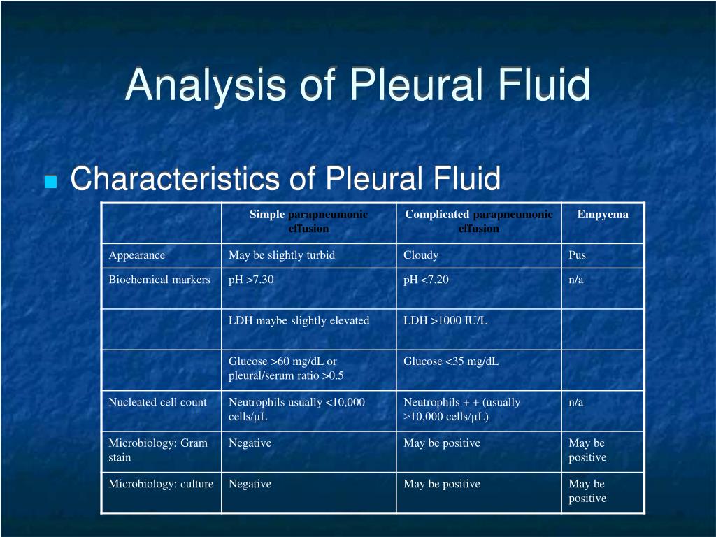
Chart Pleural Effusion Fluid Color

Color Of Pleural Effusion

Color Of Pleural Effusion
Web Various Kinds Of Fluid Can Accumulate In The Pleural Space, Such As Serous Fluid ( Hydrothorax ), Blood ( Hemothorax ), Pus ( Pyothorax, More Commonly Known As Pleural Empyema ), Chyle ( Chylothorax ), Or Very Rarely Urine ( Urinothorax) Or Feces ( Coprothorax ).
Under Normal Circumstances, A Small Amount Of Fluid Is Continuously Produced And Reabsorbed Within This Space To Maintain Lubrication And Facilitate Smooth Movement Of The Lungs During Respiration.
Web Definition (Malignant/Paramalignant) In Mpes, Infiltration Of Cancer Cells Into Pleural Tissue Is Observed;
Web Common Descriptors Were Used To Create Three Colour Charts To Describe Pleural Fluid.
Related Post: